Mandible Reconstruction with 3D Printed Models
DOI:
https://doi.org/10.5195/d3000.2024.538Parole chiave:
3D printed model, maxillofacial reconstruction, reconstruction plates, ameloblastoma, juvenile fibromatosis, keratocyst, osteosarcoma, vascular malformationAbstract
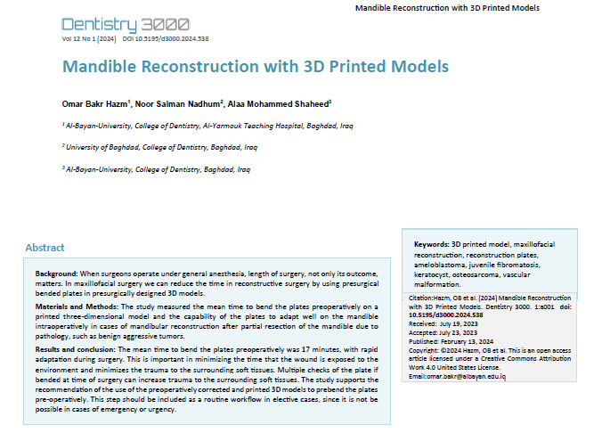
Background: When surgeons operate under general anesthesia, length of surgery, not only its outcome, matters. In maxillofacial surgery we can reduce the time in reconstructive surgery by using presurgical bended plates in presurgically designed 3D models.
Method: The study measured the mean time to bend the plates preoperatively on a printed three-dimensional model and the capability of the plates to adapt well on the mandible intraoperatively in cases of mandibular reconstruction after partial resection of the mandible due to pathology, such as benign aggressive tumors.
Results and conclusions: The mean time to bend the plates preoperatively was 17 minutes, with rapid adaptation during surgery. This is important in minimizing the time that the wound is exposed to the environment and minimizes the trauma to the surrounding soft tissues. Multiple checks of the plate if bended at time of surgery can increase trauma to the surrounding soft tissues.
The study supports the recommendation of the use of the preoperatively corrected and printed 3D models to prebend the plates pre-operatively. This step should be included as a routine workflow in elective cases, since it is not be possible in cases of emergency or urgency.
Riferimenti bibliografici
Jeroen Liebregts, Frank Baan, Pieter van Lierop, Martien de Koning, Stefaan Bergé, Thomas Maal, Tong Xi. One-year postoperative skeletal stability of 3D planned bimaxillary osteotomies: maxilla-first versus mandible-first surgery. Sci Rep. 2019 Feb 28;9(1):3000.
K Stokbro, E Aagaard, P Torkov, R B Bell, T Thygesen. Virtual planning in orthognathic surgery. Int J Oral Maxillofac Surg. 2014 Aug;43(8):957-65.
Humberto Fernández-Olarte, Andrés Gómez-Delgado, Juan G Gutiérrez-Quintero, Álvaro Rodríguez-Sáenz, Jaime Castro-Núñez. The Morpho-functional 3D Analysis for Zygomatic Implants: A Clinical Tool With Surgical Implications. J Craniofac Surg. 2020 Sep 3.
Ghada Amin Khalifa, Nahed Adly Abd El Moniem, Shadia Abd-ElHameed Elsayed, Yara Qadry. Segmental Mirroring: Does It Eliminate the Need for Intraoperative Readjustment of the Virtually Pre-Bent Reconstruction Plates and Is It Economically Valuable? J Oral Maxillofac Surg. 2016 Mar;74(3):621-30.
Fedorov A., Beichel R., Kalpathy-Cramer J., Finet J., Fillion-Robin J-C., Pujol S., Bauer C., Jennings D., Fennessy F., Sonka M., Buatti J., Aylward S.R., Miller J.V., Pieper S., Kikinis R. 3D Slicer as an Image Computing Platform for the Quantitative Imaging Network. Magnetic Resonance Imaging. 2012 Nov;30(9):1323-41. PMID: 22770690.
Azuma M, Yanagawa T, Ishibashi-Kanno N, Uchida F, Ito T, Yamagata K, Hasegawa S, Sasaki K, Adachi K, Tabuchi K, Sekido M, Bukawa H. Mandibular reconstruction using plates prebent to fit rapid prototyping 3-dimensional printing models ameliorates contour deformity. Head Face Med. 2014 Oct 23;10:45.
Cohen A, Laviv A, Berman P, Nashef R, Abu-Tair J. Mandibular reconstruction using stereolithographic 3-dimensional printing modeling technology. Oral Surg Oral Med Oral Pathol Oral Radiol Endod. 2009 Nov;108(5):661-6.
E Bradley Strong, Scott C Fuller, David F Wiley, Janina Zumbansen, M D Wilson, Marc C Metzger. Preformed vs intraoperative bending of titanium mesh for orbital reconstruction. Otolaryngol Head Neck Surg. 2013 Jul;149(1):60-6.

##submission.downloads##
Pubblicato
Fascicolo
Sezione
Licenza
Copyright (c) 2024 Omar Bakr Hazm, Noor Salman Nadhum, Alaa Mohammed Shaheed

TQuesto lavoro è fornito con la licenza Creative Commons Attribuzione 4.0 Internazionale.
Authors who publish with this journal agree to the following terms:
- The Author retains copyright in the Work, where the term “Work” shall include all digital objects that may result in subsequent electronic publication or distribution.
- Upon acceptance of the Work, the author shall grant to the Publisher the right of first publication of the Work.
- The Author shall grant to the Publisher and its agents the nonexclusive perpetual right and license to publish, archive, and make accessible the Work in whole or in part in all forms of media now or hereafter known under a Creative Commons Attribution 4.0 International License or its equivalent, which, for the avoidance of doubt, allows others to copy, distribute, and transmit the Work under the following conditions:
- Attribution—other users must attribute the Work in the manner specified by the author as indicated on the journal Web site;
- The Author is able to enter into separate, additional contractual arrangements for the nonexclusive distribution of the journal's published version of the Work (e.g., post it to an institutional repository or publish it in a book), as long as there is provided in the document an acknowledgement of its initial publication in this journal.
- Authors are permitted and encouraged to post online a prepublication manuscript (but not the Publisher’s final formatted PDF version of the Work) in institutional repositories or on their Websites prior to and during the submission process, as it can lead to productive exchanges, as well as earlier and greater citation of published work. Any such posting made before acceptance and publication of the Work shall be updated upon publication to include a reference to the Publisher-assigned DOI (Digital Object Identifier) and a link to the online abstract for the final published Work in the Journal.
- Upon Publisher’s request, the Author agrees to furnish promptly to Publisher, at the Author’s own expense, written evidence of the permissions, licenses, and consents for use of third-party material included within the Work, except as determined by Publisher to be covered by the principles of Fair Use.
- The Author represents and warrants that:
- the Work is the Author’s original work;
- the Author has not transferred, and will not transfer, exclusive rights in the Work to any third party;
- the Work is not pending review or under consideration by another publisher;
- the Work has not previously been published;
- the Work contains no misrepresentation or infringement of the Work or property of other authors or third parties; and
- the Work contains no libel, invasion of privacy, or other unlawful matter.
- The Author agrees to indemnify and hold Publisher harmless from Author’s breach of the representations and warranties contained in Paragraph 6 above, as well as any claim or proceeding relating to Publisher’s use and publication of any content contained in the Work, including third-party content.
Revised 7/16/2018. Revision Description: Removed outdated link.


