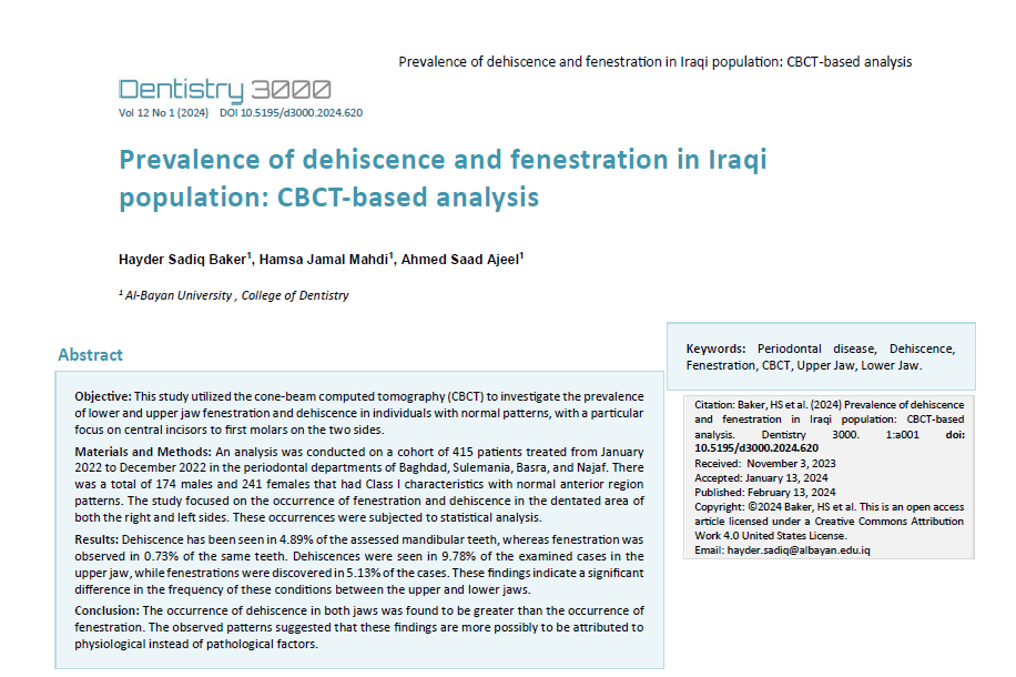
Prevalence of dehiscence and fenestration in Iraqi population: CBCT-based analysis
DOI::
https://doi.org/10.5195/d3000.2024.620کلمات کلیدی:
Periodontal disease, Dehiscence, Fenestration, CBCT, Upper Jaw, Lower Jawچکیده
Objective: This study utilized the cone-beam computed tomography (CBCT) to investigate the prevalence of lower and upper jaw fenestration and dehiscence in individuals with normal patterns, with a particular focus on central incisors to first molars on the two sides.
Materials and Methods: An analysis was conducted on a cohort of 415 patients treated from January 2022 to December 2022 in the periodontal departments of Baghdad, Sulemania, Basra, and Najaf. There was a total of 174 males and 241 females that had Class I characteristics with normal anterior region patterns. The study focused on the occurrence of fenestration and dehiscence in the dentated area of both the right and left sides. These occurrences were subjected to statistical analysis.
Results: Dehiscence has been seen in 4.89% of the assessed mandibular teeth, whereas fenestration was observed in 0.73% of the same teeth. Dehiscences were seen in 9.78% of the examined cases in the upper jaw, while fenestrations were discovered in 5.13% of the cases. These findings indicate a significant difference in the frequency of these conditions between the upper and lower jaws.
Conclusion: The occurrence of dehiscence in both jaws was found to be greater than the occurrence of fenestration. The observed patterns suggested that these findings are more possibly to be attributed to physiological instead of pathological factors.
مراجع
Accuracy and reliability of conebeam computed tomography for measuring alveolar bone height and detecting bony dehiscences and fenestrations. Leung CC, Palomo L, Griffith R, Hans MG. Am J Orthod Dentofac Orthop. 2010;137(4 Suppl):S109–19.
Periodontal condition in orthodontically treated and untreated individuals II. Alveolar bone losss: radiographic findings. Zachrisson BU, Alnaes L. Angle Orthod. 1974;44(1):48–55.
Trabecular and cortical bone as risk factors for orthodontic relapse. Rothe LE, Bollen A-M, Little RM, Herring SW, Chaison JB, Chen CSK, et al. Am J Orthod Dentofac Orthop. 2006;130(4):476–84.
Mucogingival considerations in orthodontic treatment. Wennström JL. Semin Orthod. 1996;2(1):46–54.
Evaluation of dehiscence and fenestration in adolescent patients affected by unilateral cleft lip and palate: a retrospective cone beam computed tomography study. Buyuk SK, Ercan E, Celikoglu M, Sekerci AE, Hatipoglu M. Angle Orthod. 2016;86(3):431–6.
Evaluation of alveolar bone loss following rapid maxillary expansion using cone-beam computed tomography. Baysal A, Uysal T, Veli I, Ozer T, Karadede I, Hekimoglu S. Korean J Orthod. 2013;43(2):83–95.
Alveolar bone changes after asymmetric rapid maxillary expansion. Akin M, Baka ZM, Ileri Z, Basciftci FA. Angle Orthod. 2015;85(5):799–805.
Dehiscence and fenestration in patients with Class I and Class II Division 1 malocclusion assessed with cone-beam computed tomography. Evangelista K, Vasconcelos Kde F, Bumann A, Hirsch E, Nitka M, Silva MA. Am J Orthod Dentofac Orthop. 2010;138(2):133.e1–7.
Dehiscence and fenestration in patients with different vertical growth patterns assessed with cone-beam computed tomography. Enhos S, Uysal T, Yagci A, Veli I, Ucar FI, Ozer T. Angle Orthod. 2012;82(5):868–74.
Dehiscence and fenestration in skeletal Class I, II, and III malocclusions assessed with cone-beam computed tomography. Yagci A, Veli I, Uysal T, Ucar FI, Ozer T, Enhos S. Angle Orthod. 2012;82(1):67–74.
Carranza FA. Carranza’s clinical periodontology. Newman MG, Takei HH, Klokkevold PR, 12th ed. St. Louis, MO: Elsevier; 2015.
Appraisal of the relationship between tooth inclination, dehiscence, fenestration, and sagittal skeletal pattern with cone beam computed tomography. Coşkun İ, Kaya B. The Angle Orthodontist. 2019;89(4):544–51.
Alveolar defects in human skulls. Davies RM, Downer MC, Hull PS, Lennon MA J Clin Periodontol. 1974; 1:107–111
Alveolar bone dehiscences and fenestrations: an anatomical study and review. Nimigean VR, Nimigean V, Bencze MA, Dimcevici-Poesina N, Cergan R, Moraru S. Rom J Morphol Embryol. 2009; 50(3):391–397
Alveolar plate fenestrations and dehiscences of the human skull. Larato DC (1970) Oral Surg Oral Med Oral Pathol. 1970; 29:816–819
Alveolar plate defects in children’s skulls. Larato DC. J Periodontol, 1972 43:502.
Prevalence of dehiscences and fenestrations in modern American skulls. Rupprecht RD, Horning GM, Nicoll BK, Cohen ME. J Periodontol. 2001; 72:722–729.
Incidence and distribution of alveolar bony dehiscence and fenestration in dry human Egyptian jaws. Abdelmalek RG, Bissada NF. J Periodontol. 1973; 44:586–588.
Bony defects in dried Bantu mandibles. Volchansky A, Cleaton-Jones P. Oral Surg Oral Med Oral Pathol. 1978; 45:647–653.
Alveolar bone fenestrations and dehiscences in dry Bedouin jaws. Edel A. J Clin Periodontol. 1981; 8:491–499.
The correlation between the presence of dehiscence or fenestration and the severity of tooth attrition in contemporary dry Japanese adult skulls–part I. Ezawa T, Sano H, Kaneko K, Hiruma S, Fujikawa K, Murai S. J Nihon Univ Sch Dent. 1987; 29:27–34
Dehiscence and fenestration: study of distribution and incidence in a homogeneous population model. Urbani G, Lombardo G, Filippini P, Nocini FP. Stomatol Mediterr, 1991; 11:113–118
Dehiscence and fenestration in patients with Class I and Class II Division 1 malocclusion assessed with conebeam computed tomography. Evangelista K, Vasconcelos KDF, Bumann A, Hirsch E, Nitka M, Silva MAG. Am J Orthod Dentofacial Orthop. 2010; 138:131–133
Depth of alveolar bone dehiscences in relation to gingival recessions. Lost C. J Clin Periodontol. 1984; 11:583–589.
Evaluation of dehiscence and fenestration in adolescent patients affected by unilateral cleft lip and palate: a retrospective cone beam computed tomography study. Buyuk SK, Ercan E, Celikoglu M, Sekerci AE, Hatipoglu M. Angle Orthod. 2016; 86:431–436.
The effect of orthodontic treatment on periodontal bone support in patients with advanced loss of marginal periodontium. Artun J, Urbye KS. Am J Orthod Dentofacial Orthop. 1988; 93:143–148.
Periodontal tissue response to orthodontic movement of teeth with infrabony pockets. Wennstrom JL, Stokland BL, Nyman S, Thilander B. Am J Orthod Dentofacial Orthop. 1993; 103:313–319.
Accuracy and reliability of buccal bone height and thickness measurements from cone-beam computed tomography imaging. Timock AM, Cook V, McDonald T, Leo MC, Crowe J, Benninger BL et al. Am J Orthod Dentofacial Orthop. 2011; 140:734–744.
Accuracy and reliability of cone-beam computed tomography for measuring alveolar bone height and detecting bony dehiscences and fenestrations. Leung CC, Palomo L, Griffith R, Hans MG. Am J Orthod Dentofacial Orthop. 2010; 137(4 Suppl):109–119
Accuracy of cone-beam computed tomography in detecting alveolar bone dehiscences and fenestrations. Sun L, Zhang L, Shen G, Wang B, Fang B. Am J Orthod Dentofacial Orthop. 2015; 147:313–323
Accuracy of alveolar bone measurements from cone beam computed tomography acquired using varying settings. Cook VC, Timock AM, Crowe JJ, Wang M, Covell DA Jr. Orthod Craniofacial Res. 2015;18(S1):127–36.
Changes of alveolar bone dehiscence and fenestration after augmented corticotomy-assisted orthodontic treatment: a CBCT evaluation. Sun L, Yuan L, Wang B, Zhang L, Shen G, Fang B. Prog Orthod. 2019; 20:7.
Clinical recommendations regarding use of cone beam computed tomography in orthodontics.[corrected]. Position statement by the American Academy of Oral and Maxillofacial Radiology. American Academy of O, Maxillofacial R. Oral Surg Oral Med Oral Pathol Oral Radiol. 2013; 116(2):238–57.
The prevalence of dehiscence and fenestration on anterior region of skeletal Class III malocclusions: a cone-beam CT study. Sun LY, Wang B, Fang B. Shanghai Kou Qiang Yi Xue. 2013; 22:418–422
Evaluation of alveolar bone defects on anterior region in patients with bimaxillary protrusion by using cone-beam CT. Zhou L, Li WR. Beijing Da Xue Xue Bao Yi Xue Ban. 2015; 47:514–520
Prevalence of posterior alveolar bony dehiscence and fenestration in adults with posterior crossbite: a CBCT study. Choi JY, Chaudhry K, Parks E, Ahn JH. Prog Orthod. 2020; 21:8
Incidence and distribution of dehiscences and fenestrations on human skulls. Jorgic-Srdjak K, Plancak D, Bosnjak A, Azinovic Z. Coll Antropol. 1998; 22(Suppl):111–116.
Accuracy of cone-beam computed tomography in detecting alveolar bone dehiscences and fenestrations. Sun L, Zhang L, Shen G, Wang B, Fang B. Am J Orthod Dentofac Orthop. 2015;147(3):313–23.
Upper incisor position and bony support in untreated patietns as seen on CBCT. Gracco A, Lombardo L, Mancuso G, Gravina V, Siciliani G. Angle Orthodontist. 2009;79(4):692–702.

فایلهای دیگر
چاپ شده
شماره
نوع مقاله
مجوز
حق نشر 2024 Hayder Sadiq Baker, Hamsa Jamal Mahdi, Ahmed Saad Ajeel

این پروژه تحت مجوز بین المللی Creative Commons Attribution 4.0 می باشد.
Authors who publish with this journal agree to the following terms:
- The Author retains copyright in the Work, where the term “Work” shall include all digital objects that may result in subsequent electronic publication or distribution.
- Upon acceptance of the Work, the author shall grant to the Publisher the right of first publication of the Work.
- The Author shall grant to the Publisher and its agents the nonexclusive perpetual right and license to publish, archive, and make accessible the Work in whole or in part in all forms of media now or hereafter known under a Creative Commons Attribution 4.0 International License or its equivalent, which, for the avoidance of doubt, allows others to copy, distribute, and transmit the Work under the following conditions:
- Attribution—other users must attribute the Work in the manner specified by the author as indicated on the journal Web site;
- The Author is able to enter into separate, additional contractual arrangements for the nonexclusive distribution of the journal's published version of the Work (e.g., post it to an institutional repository or publish it in a book), as long as there is provided in the document an acknowledgement of its initial publication in this journal.
- Authors are permitted and encouraged to post online a prepublication manuscript (but not the Publisher’s final formatted PDF version of the Work) in institutional repositories or on their Websites prior to and during the submission process, as it can lead to productive exchanges, as well as earlier and greater citation of published work. Any such posting made before acceptance and publication of the Work shall be updated upon publication to include a reference to the Publisher-assigned DOI (Digital Object Identifier) and a link to the online abstract for the final published Work in the Journal.
- Upon Publisher’s request, the Author agrees to furnish promptly to Publisher, at the Author’s own expense, written evidence of the permissions, licenses, and consents for use of third-party material included within the Work, except as determined by Publisher to be covered by the principles of Fair Use.
- The Author represents and warrants that:
- the Work is the Author’s original work;
- the Author has not transferred, and will not transfer, exclusive rights in the Work to any third party;
- the Work is not pending review or under consideration by another publisher;
- the Work has not previously been published;
- the Work contains no misrepresentation or infringement of the Work or property of other authors or third parties; and
- the Work contains no libel, invasion of privacy, or other unlawful matter.
- The Author agrees to indemnify and hold Publisher harmless from Author’s breach of the representations and warranties contained in Paragraph 6 above, as well as any claim or proceeding relating to Publisher’s use and publication of any content contained in the Work, including third-party content.
Revised 7/16/2018. Revision Description: Removed outdated link.


